Drawing from the Past
Celebrating the fine art of science illustration
For more than 75 years, the Temerty Faculty of Medicine’s biomedical communications (BMC) program has married art and science to help physicians and later the general public understand the world in new and beautiful ways.
Founded in 1945, BMC is the only Canadian program — and one of just five in North America — to offer highly specialized graduate-level training to develop visual material for health promotion, medical education and as part of the process of scientific discovery.
Beginning as the Art as Applied to Medicine (AAM) Certificate Program, it became a bachelor of science degree (BScAAM) in the 1960s. Then in the 1990s, the program evolved again with a new name — biomedical communications — and a new mandate: to offer instruction at the graduate level.
Over the decades, the illustrators’ art supplies have advanced as quickly as the scientific and medical subjects they depict.
In the early days, program founder Maria Torrence Wishart and contemporaries Dorothy Foster Chubb and Elizabeth Blackstock used carbon dust, pen and ink, and paint to create anatomical illustrations for medical textbooks and teaching materials. By the latter half of the 20th century, photography, video and 35mm slides added new instruments to the communicators’ tool box, and their work began to reach lay audiences.
Today, biomedical illustrators rely on advanced technology to communicate more complex subjects. Digital animation and design platforms are now the go-to tools for illustration, while gaming systems and augmented reality programs are at the forefront for animations and surgical technique simulations.
Advances in science and medicine have also influenced the field, offering new opportunities to showcase discoveries at the cellular or even molecular level, as seen in the illustrations by BMC alumni Naveen Devasagayam and Sam Holmes that accompany this story.
The work of biomedical communicators has found a broader audience, too. Once aimed exclusively at scientists and health care professionals, medical illustration and animation now appear in medical advertising, educational gaming, public health campaigns and nightly news stories.
-
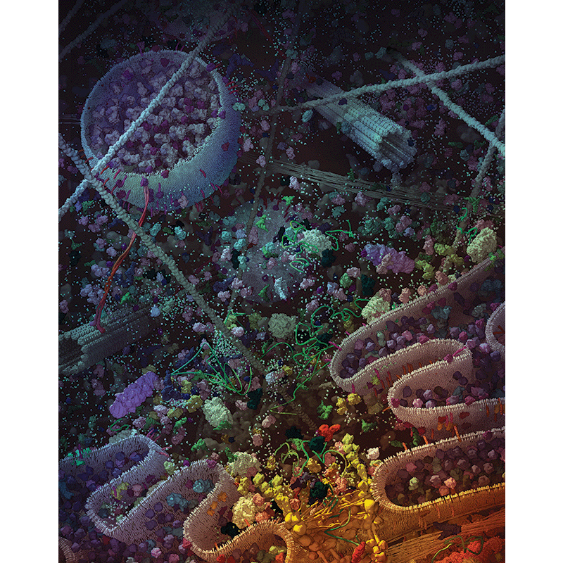 “Portrait of a cell,” 2015 by Naveen Devasagayam (MScBMC ’15). Contemporary medical illustrators combine scientific knowledge and design expertise to bring biomedical data to life. In this image, Devasagayam demonstrates the variety of molecular populations inside a eukaryotic cell.
 -
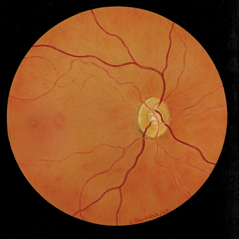
“Retina,” 1943 by Elizabeth Blackstock (DSAM ’47). Before it was possible to obtain high-quality photographs of the retina, medical illustrators used watercolours or gouache to capture the nuances of retinal form, texture and colour.
-
“Encoding Nature’s Chemicals,” still from digital animation, 2016 by Sam Holmes (MScBMC ’15). Holmes created a digital animation about the search for novel therapeutics within the DNA of microbes in the environment. -
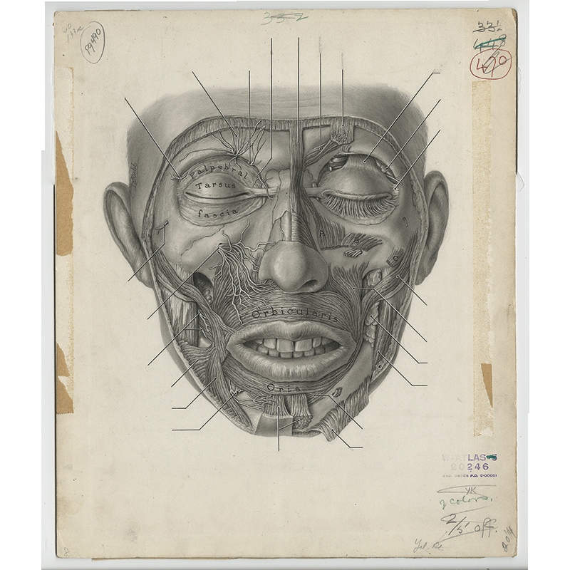
“Facial anatomy,” 1943 by Dorothy Foster Chubb. Chubb was a member of a small team of women who illustrated Grant’s Atlas of Anatomy, produced at the University of Toronto and now in its 15th edition. This carbon dust illustration bears the marks of its journey through the publishing process. -
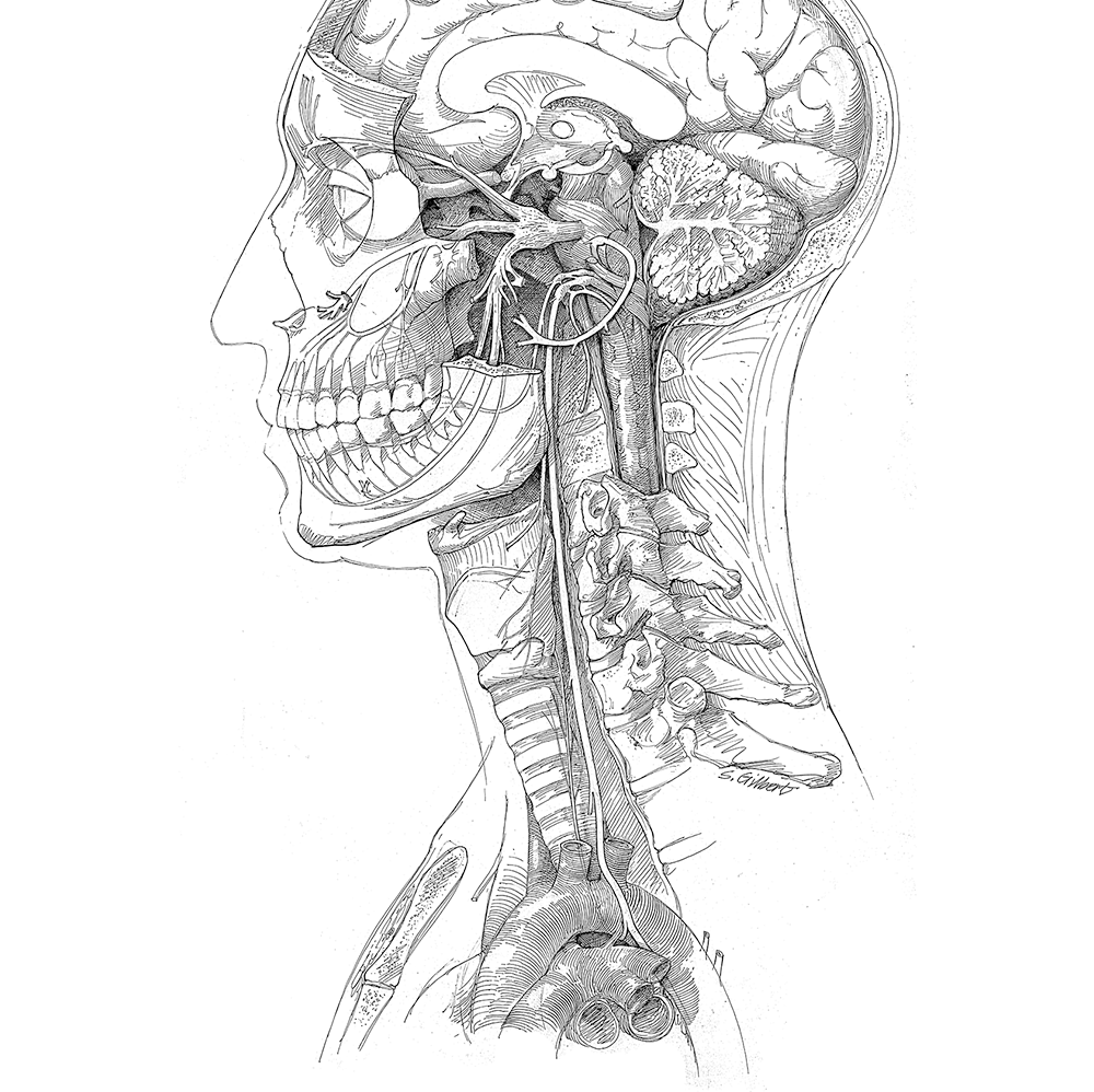
“Cranial nerves,” undated by Stephen Goltra Gilbert, a long-time faculty member in Biomedical Communications, and a master of pen-and-ink. This technique was a staple for medical illustrators before the digital age. -
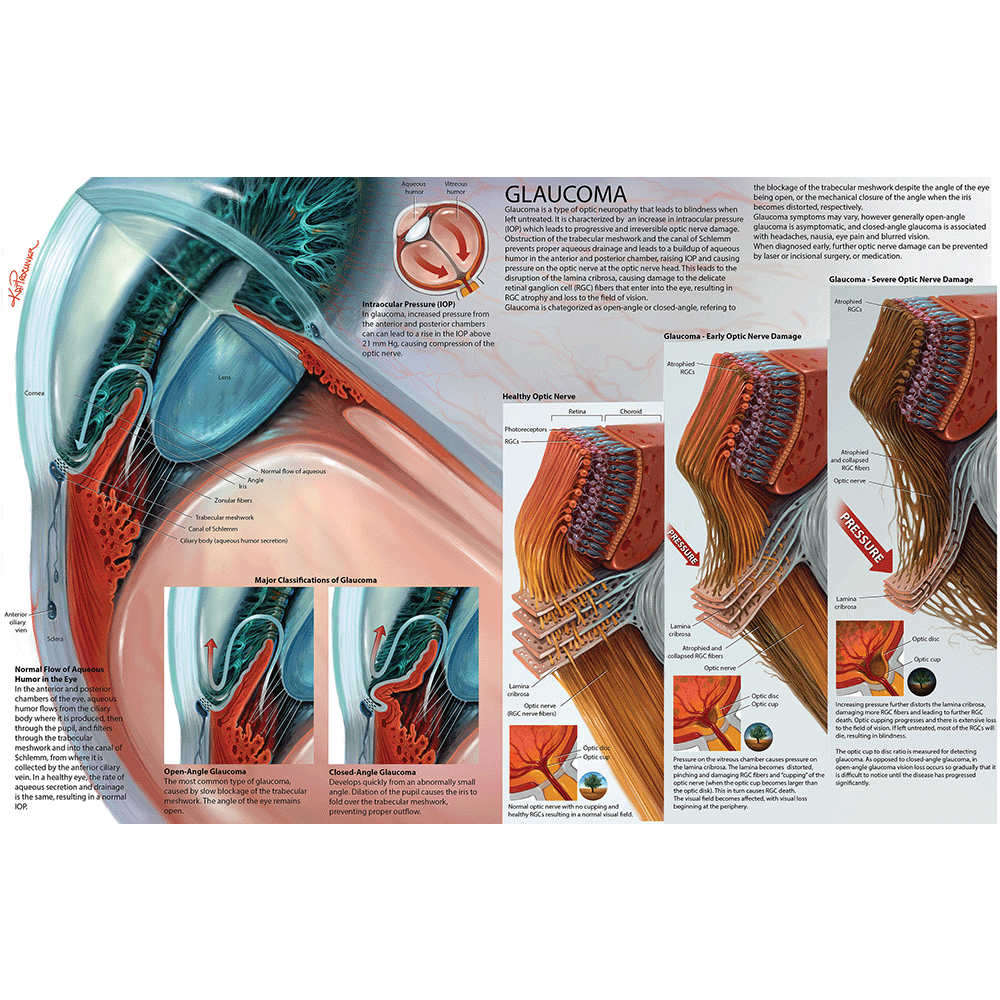
“Glaucoma,” 2014 by Kateryna Procunier (MScBMC ’15). In this digital illustration, alumna Procunier depicts the cause and progression of glaucoma and its effects on the visual field. -
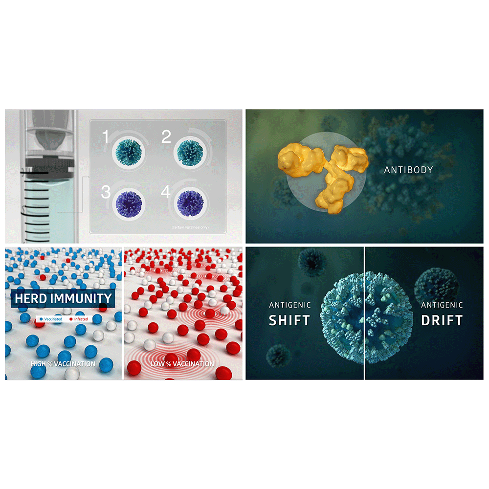
“Flu Facts,” (animation stills), 2015 by Natalie Cormier (MScBMC ’15). Animation now plays a large role in biomedical visualization. Cormier created the 3D animation as a Master’s student in the BMC program, to explain the importance of seasonal vaccination. -
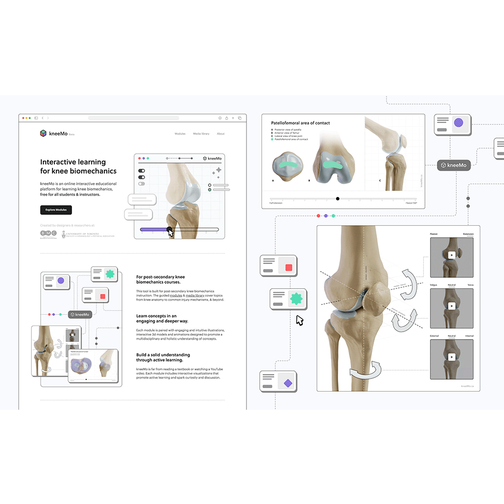
“KneeMo,” app screen captures, 2022 by Shehryar (Shay) Saharan (MScBMC ’22). Along with animation, interactive media is now an important tool in biomedical visualization. KneeMo, created as a BMC Master’s research project, is an online, interactive, educational platform for learning knee biomechanics. -
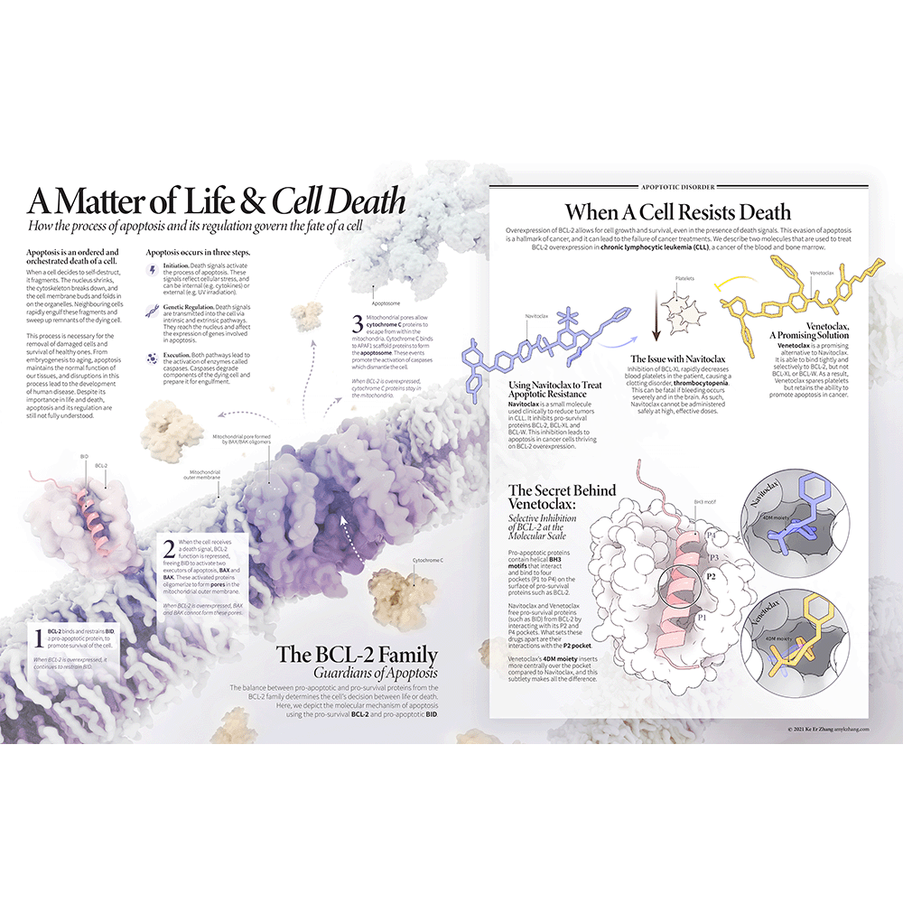
Amy Ke Er Zhang, “A Matter of Life & Cell Death,” 2021 by Amy Ke Er Zhang (MScBMC ’22). Zhang uses molecular data and digital rendering to explain the process of cell death and the mechanism of action of two molecules used in the treatment of chronic lymphocytic leukemia.